- 50% SALE
- Anatomy of the Eye
- Digital Acuity
- Domiciliary
- Illuminated Cabinets
- Low Vision Aids
- New Products
- Charts & Distance Tests
- COVID
- Clinical Trials Products
- Dispensing & Workshop
- Field Screening
- General Refraction
- Amsler Chart
- Autorefractors & Keratometers
- Bagolini Striated Lenses
- Cardiff Cards
- Clip-on Occluder
- Colour Tests
- Cross Cylinders
- Domiciliary
- Filter Bars
- Fixation Sticks
- Halberg Clips
- Lens Confirmation Tests
- Maddox Phoria Measure
- Maddox Wing Test
- Near Vision
- Occluders
- Optician Starter Kit
- Optokinetic Drum
- Paddle Retinoscopy Rack Set
- Pen Torch
- Polarising Visor
- Prisms & Prism Bars
- Refracto-Rack
- Stereo Tests
- Trial Frames
- Trial Lens Sets
- Trial Lens Spares
- Volk & Ocular Lenses
- Furniture
- Scopes & Loupes
- Sports Vision
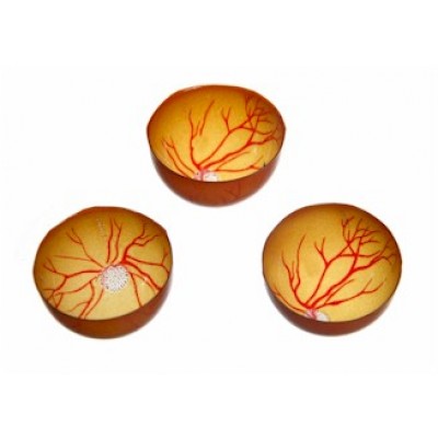
Reti Eye Laser Practice Retinal Films 10 Pack
These films are compatible with the Reti Eye Laser Practice Kit.
There are 10 replacement films per pack.
The Reti Eye Laser Practice Kit has a 6mm pupil and is comprised of two halves – the upper half includes a lens; the lower half houses replaceable retinal films that show vascularization and the optic nerve.
The kit includes the Reti Eye, a wooden base, 10 retinal films, and an adjustable clamp for slit lamp mounting.
For indirect ophthalmoscopy practice, the Reti Eye is placed on the wood base and rotated for a clear view of the fundus.
During photocoagulation practice, tiny white spots form on the retinal film when using a green photocoagulation laser.


